Services
Ultrasound
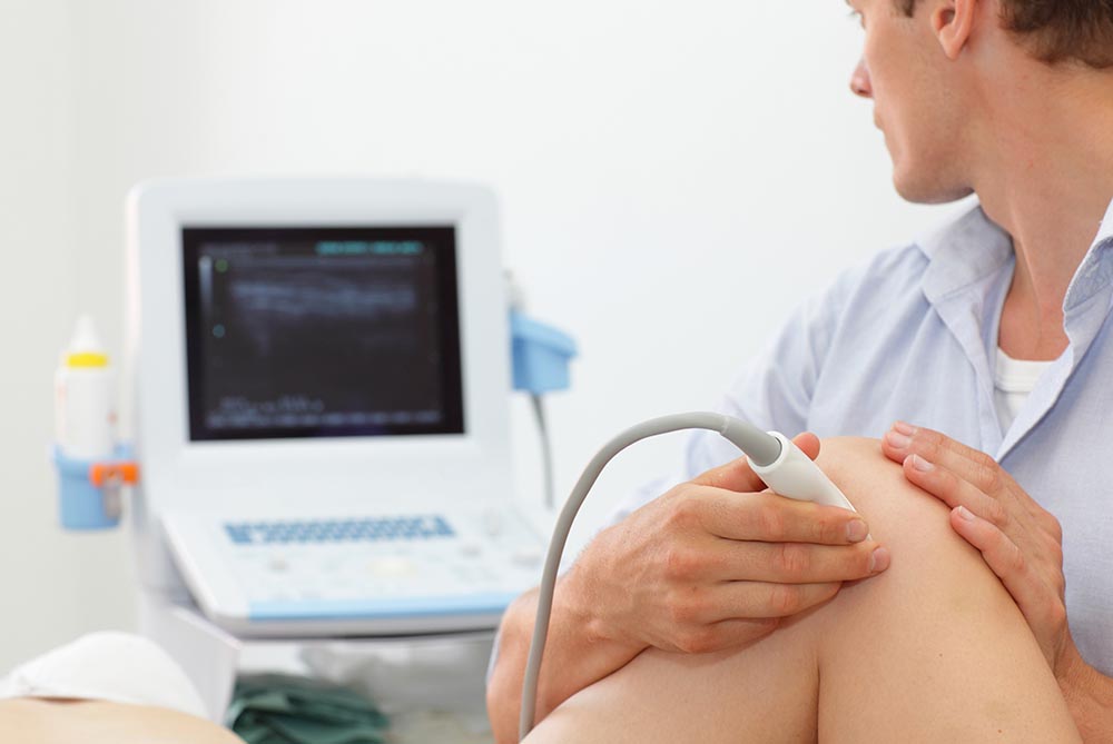
An ultrasound is a procedure in which high-energy sound waves are bounced off internal tissues or organs and make echoes. The echo patterns are shown on the screen of an ultrasound machine, forming a picture of body tissues called a sonogram.
Exam Preparation
These instructions are IMPORTANT. Please follow them.
ABDOMEN
Includes studies of the gall bladder, pancreas, spleen, liver, kidneys and aorta. If your appointment is in the morning, DO NOT eat or drink anything 12 hours prior to your examination. If your appointment is in the afternoon, for breakfast you may eat dry toast, black tea, black coffee, juice up to 9 a.m. Nothing to drink or eat after that. These instructions are important as we require you to have an empty stomach.
PELVIS
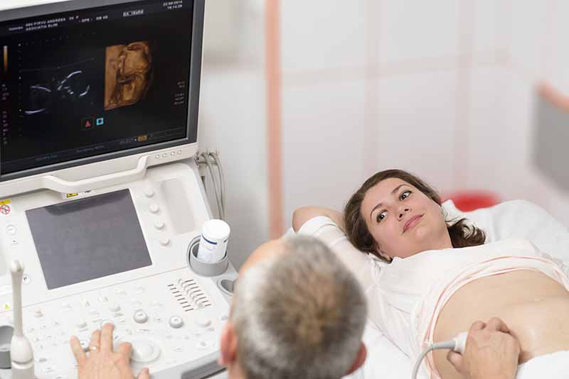
Includes transvaginal, uterus, ovaries, bladder, g.u.tract and pregnancy (obstetrical/nuchal translucency).
You must drink 32 ounces or 1 litre (4 large glasses) of fluids completed 1 hour prior to your appointment. This can include coffee, tea, juice, water, etc. – not milk. Do not go to the washroom. You must have a full bladder for this examination. We will try to examine you as soon as possible on arrival so that you will not have to be uncomfortable for too long. Eat the meal nearest your examination – there is no reason not to eat.
ABDOMEN & PELVIS
DO NOT eat anything 12 hours prior to examination. Finish drinking 32 ounces (1 litre) of water, and ONLY water one hour before your examination. Do not go to the washroom.
PROSTATE WITH TRANSRECTAL
Finish drinking 32 ounces or 1 litre (4 large glasses) of water 1 hour before appointment. Do not go to the washroom. Take mild laxative the evening before. (PROSTATE ONLY – OMIT LAXATIVE).
THYROID
No preparation. May be booked at any time during the day/evening.
SCROTAL
No preparation. May be booked at any time during the day/evening.
BREAST
No preparation. May be booked at any time during the day/evening.


Vascular Ultrasound
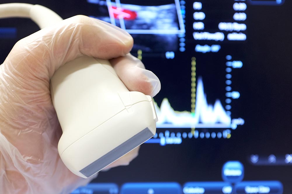
Vascular ultrasound can be used to evaluate: The blood flow in the arteries in your neck that supply blood to the brain. The blood flow to a newly transplanted organ. Blood flow in the arteries to detect the presence, severity and specific location of a narrowed area of the arteries
Why is a Vascular Ultrasound Done?
Vascular ultrasound is performed to: help monitor the blood flow to organs and tissues throughout the body. locate and identify blockages (stenosis) and abnormalities like plaque or emboli and help plan for their effective treatment.

Digital X-Ray
X-ray is the oldest and most frequently used form of medical imaging. It is the fastest and easiest way for a physician to view and assess bone fractures, cancer, pneumonia, spinal problems, blocked arteries and other abnormalities. These exams take seconds to perform and are painless.
Often multiple images are taken from different angles so a more complete view of the area is available.
Digital X-rays use small amounts of radiation. The level of exposure is considered safe for adults. However, it is not considered safe for a developing fetus. Be sure to tell the X-ray technician before the procedure if you are pregnant or think you may be pregnant. Your doctor may suggest a different testing method that does not use radiation.
The images are then read by our radiologist, who is a physician with specialized training in X-ray and other imaging tests, and a report will be sent to your doctor in a timely manner.
Why an X-Ray Is Performed
Your doctor may order an X-ray if he or she needs to look inside your body. For example, your doctor may want to:
- view an area where you are experiencing pain
- monitor the progression of a disease, such as osteoporosis
- see the effect of a treatment method
- rule out possibility of a broken bone
How to Prepare for an X-Ray
X-rays are standard procedures and involve almost no preparation from the patient.
Depending on the area under review, you may want to wear loose, comfortable clothing that you can easily move around in. You may also be asked to change into a gown for the test.
You will be instructed to remove any jewelry and other metallic items from your body before the X-ray is taken. You should let the X-ray technician know if you have any metal implants from prior surgeries. These can block the X-rays from passing through your body.

Bone Mineral Density
A bone mineral density (BMD) test uses X-rays to measure how much calcium and other types of minerals are in your bones.
This test is important for people who are at risk for osteoporosis, especially women and older adults. It also helps your health care provider detect osteoporosis and predict your risk of bone fractures.
How the Test Will Feel
The scan is safe, painless and requires no medication. You simply need to lie on a table and remain still during the test. An X-ray machine scans your hip, spine, and other bones of your torso.
Why the Test is Performed
Bone mineral density (BMD) tests are used to:
- Diagnose bone loss and osteoporosis
- See how well osteoporosis medicine is working
- Predict your risk of future bone fractures
Indications for BMD testing
may include one or more of the following:
- Age 65 or older – men and women
- Fragility fracture after age 40
- Vertebral fracture or low bone mass identified on X-ray imaging
- Parental hip fracture
- High alcohol intake
- Current smoking
- Low body weight, i.e. less than 132 lbs or 60 kg
- Rheumatoid arthritis
- Use of corticosteroid drugs (for three months or more)

